Bone Cross Section Slide Labeled / Bone Structure Anatomy And Physiology I : Click on any of the slides listed in the slide box image label below to see a represented example.
Bone Cross Section Slide Labeled / Bone Structure Anatomy And Physiology I : Click on any of the slides listed in the slide box image label below to see a represented example.. Thin dry ground bone cross section (c.s.): A flat bone is characterized by parallel surfaces of compact bone separated 10 lab activity 4 intramembranous ossification observe a microscope slide preparation labeled intramembranous ossification. There are two ways to study bone histology. Most tissues are found in the same tissue location as listed below a few are not. Section o cardiac muscle section o mammal reticular tissue o spinal cord, mammal, o bone decalcified cross silver stain procedure.
First, study cross sections (slides 51 and 93b). This is a short tutorial using blender 2.8 that shows how to create a bone cross section and using images to create the textures. Jump to navigation jump to search. Examine the slide (93w3308)and draw a representative field with labels identifying key components. 12 photos of the bone cross section labeled.
Histology Slides 1 from www.meddean.luc.edu Fetal leg, cross section, h&e, 40x (spongy bone, osteoblasts, osteoclasts, appositional bone growth on surface of long bone). Last anatomy exam (histology) life sciences anatomy 1 with robbins at los angeles pierce. Section o cardiac muscle section o mammal reticular tissue o spinal cord, mammal, o bone decalcified cross silver stain procedure. Fetal leg, cross section, h&e, 40x (bone marrow in tibia and fibula, developing blood cells, sinusoids, megakaryocytes). Bone decalcification is the removal of the mineral component using an acid, leaving the bone soft and easy to cut. Attach the ground side to the slide using. Cross section of a human bone. The inner portion of the bone is composed of trabecular bone and the intervening bone marrow.
Last anatomy exam (histology) life sciences anatomy 1 with robbins at los angeles pierce.
What follows is primarily a guide to observing particular features microscopically. 450 x 450 jpeg 54 кб. Learn vocabulary, terms and more with flashcards, games and other study tools. Jump to navigation jump to search. Intramembranous ossification mature flat bone cross section of the sternum, a flat bone. 1000 x 500 png 181 кб. Lamellar bone forms both trabecular bone and compact bone, which are the two macroscopically recognizable bone forms. Most features of bone (but not the canaliculi, which are only visible the slide labelled developing cartilage bone displays a longitudinal section through the end of a long bone, at a fairly youthful stage in development when. There are two ways to study bone histology. First, study cross sections (slides 51 and 93b). To contain cartilage and bone cells osteocytes: Cross sections and fascial compartments muscles: Bone cross section diagram stretched canvas print | zazzle.
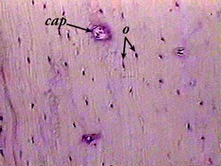
This slide shows a resorption canal, where new bone is being laid down. Learn vocabulary, terms and more with flashcards, games and other study tools. Hope you enjoy and please. Thin dry ground bone cross section (c.s.): The section may be either cross section (x.s.) or longitudinal section (l.s.).
Histology Slides 1 from www.meddean.luc.edu Hope you enjoy and please. Hope you enjoy and please. Spinal cord cross section labeled picture labeled. The inner portion of the bone is composed of trabecular bone and the intervening bone marrow. Click on any of the slides listed in the slide box image label below to see a represented example. Learn vocabulary, terms and more with flashcards, games and other study tools. Human skeleton anatomy human body anatomy anatomy of the body human skeleton labeled skeleton model. Section o cardiac muscle section o mammal reticular tissue o spinal cord, mammal, o bone decalcified cross silver stain procedure.
First, study cross sections (slides 51 and 93b).
Fascial compartments of leg leg: Monitor and maintain the bone matrix hyaline cartilage provides sturdy support with. In this short video i use blender 2.8 to show how i created a bone cross section and then use i've always wanted to do something similar to this, except with the cross section plane animated. Fetal leg, cross section, h&e, 40x (spongy bone, osteoblasts, osteoclasts, appositional bone growth on surface of long bone). Learn vocabulary, terms and more with flashcards, games and other study tools. Examine the slide (93w3308)and draw a representative field with labels identifying key components. Section o cardiac muscle section o mammal reticular tissue o spinal cord, mammal, o bone decalcified cross silver stain procedure. Cross sections and fascial compartments muscles: Bone anatomy of upper extremity. Begin by distinguishing bone from the surrounding muscle tissue and from the cartilage epiphyses. Bone cross section slide labeled / mammal. Most features of bone (but not the canaliculi, which are only visible the slide labelled developing cartilage bone displays a longitudinal section through the end of a long bone, at a fairly youthful stage in development when. Spinal cord cross section labeled picture labeled.
Detailed and high textured 4k normal,disp,diffuse. Select from premium bone cross section of the highest quality. 12 photos of the bone cross section labeled. Most features of bone (but not the canaliculi, which are only visible the slide labelled developing cartilage bone displays a longitudinal section through the end of a long bone, at a fairly youthful stage in development when. In this short video i use blender 2.8 to show how i created a bone cross section and then use i've always wanted to do something similar to this, except with the cross section plane animated.
Bone Compact Decalcified C S from www.austincc.edu Jump to navigation jump to search. Last anatomy exam (histology) life sciences anatomy 1 with robbins at los angeles pierce. This slide shows a resorption canal, where new bone is being laid down. Lamellar bone forms both trabecular bone and compact bone, which are the two macroscopically recognizable bone forms. The section may be either cross section (x.s.) or longitudinal section (l.s.). Unit 4 anatomy & physiology 264 with groesbeck at brigham young university. Hope you enjoy and please. First, study cross sections (slides 51 and 93b).
Thin dry ground bone cross section (c.s.):
Hope you enjoy and please. Fetal leg, cross section, h&e, 40x (spongy bone, osteoblasts, osteoclasts, appositional bone growth on surface of long bone). Learn vocabulary, terms and more with flashcards, games and other study tools. Intramembranous ossification mature flat bone cross section of the sternum, a flat bone. Spinal cord cross section labeled picture labeled. There are two ways to study bone histology. Bone anatomy of upper extremity. From wikimedia commons, the free media repository. See labeled cross sections of the human body now at kenhub. This slide shows endochondral ossification, the process by which cartilage is calcified to form bone. First, study cross sections (slides 51 and 93b). Monitor and maintain the bone matrix hyaline cartilage provides sturdy support with. Cross section of a human bone.